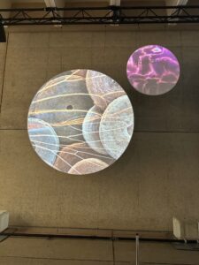
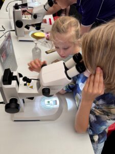
The World Science Festival’s Unseen Worlds exhibition, was held in the Whale Mall at Queensland Museum over the weekend. The event captivated over 1000 visitors of all ages with its intricate displays and interactive experiences, showcasing a stunning array of microscopic wonders, from bugs and natural items to quantum chips. Visitors had the opportunity to explore these hidden micro-worlds through ten Zeiss microscopes, revealing microscopic mysteries that are usually invisible to the naked eye.
The exhibition featured a visual projection with images curated by some of Queensland’s leading scientific researchers and organisations, including QBiotics Group; donna davis – artist-in-residence, Queensland Herbarium and Biodiversity Science Unit; Geoff Thompson and Lily Kumpe, Queensland Museum; The University of Queensland’s Institute for Molecular Bioscience (IMB) and QUBIC; with soundscape design by Luke Lickfold. Attendees were thrilled by the detailed displays and the chance to engage with the science that shapes our world. The interactive microscopy activation allowed participants to dive deep into the microscopic universe, sparking curiosity and wonder among both young and old.
The Unseen Worlds exhibition not only highlighted the beauty and complexity of the microscopic world but also underscored the importance of scientific research and innovation.
QUBIC is excited to announce the addition of Emma De Costa to the team. Emma has started on her PhD journey in our Brain Theme under the mentorship of Prof. Lezanne Ooi and Prof. Haibo Yu, in collaboration with Prof. Youngchan Kim and Prof. Marco Sacchi, from the University of Surrey. Emma is the recipient of the University of Wollongong / University of Surrey Joint-Dual Degree PhD Scholarship.
Her research focuses on TDP-43, a protein strongly linked to Amyotrophic Lateral Sclerosis (ALS). Emma aims to investigate how disease and structure-linked mutations affect TDP-43 aggregation kinetics and phase separation behaviour, using novel quantum tools as well as techniques like Brillouin light scattering microscopy.
“Traditional biophysical methods often lack the sensitivity and resolution to capture real-time processes.” says Emma. “By collaborating across QUBIC teams, we’re using quantum sensing techniques, and Brillouin light scattering microscopy to overcome these challenges. This will help us understand how specific mutations affect TDP-43 dysfunction and contribute to neurodegenerative diseases.”
Emma’s path to science was unconventional. Initially enrolled in Science Education, she discovered her true passion lay in the science itself, so she switched to a Bachelor of Science, where she completed her Honours year researching Parkinson’s disease.
Emma finds immense reward in the collaborative and communicative aspects of science. She values the supportive community at the University of Wollongong where she’s based. Her colleagues’ humour and dedication make every day in the lab worthwhile.
A strong advocate for diversity in science, Emma encourages young people to pursue their interests regardless of background or challenges. She believes science offers diverse opportunities, from technology and coding to fieldwork and data analytics.
QUBIC is thrilled to have Emma on board, and we look forward to her contributions to neurodegenerative disease research!
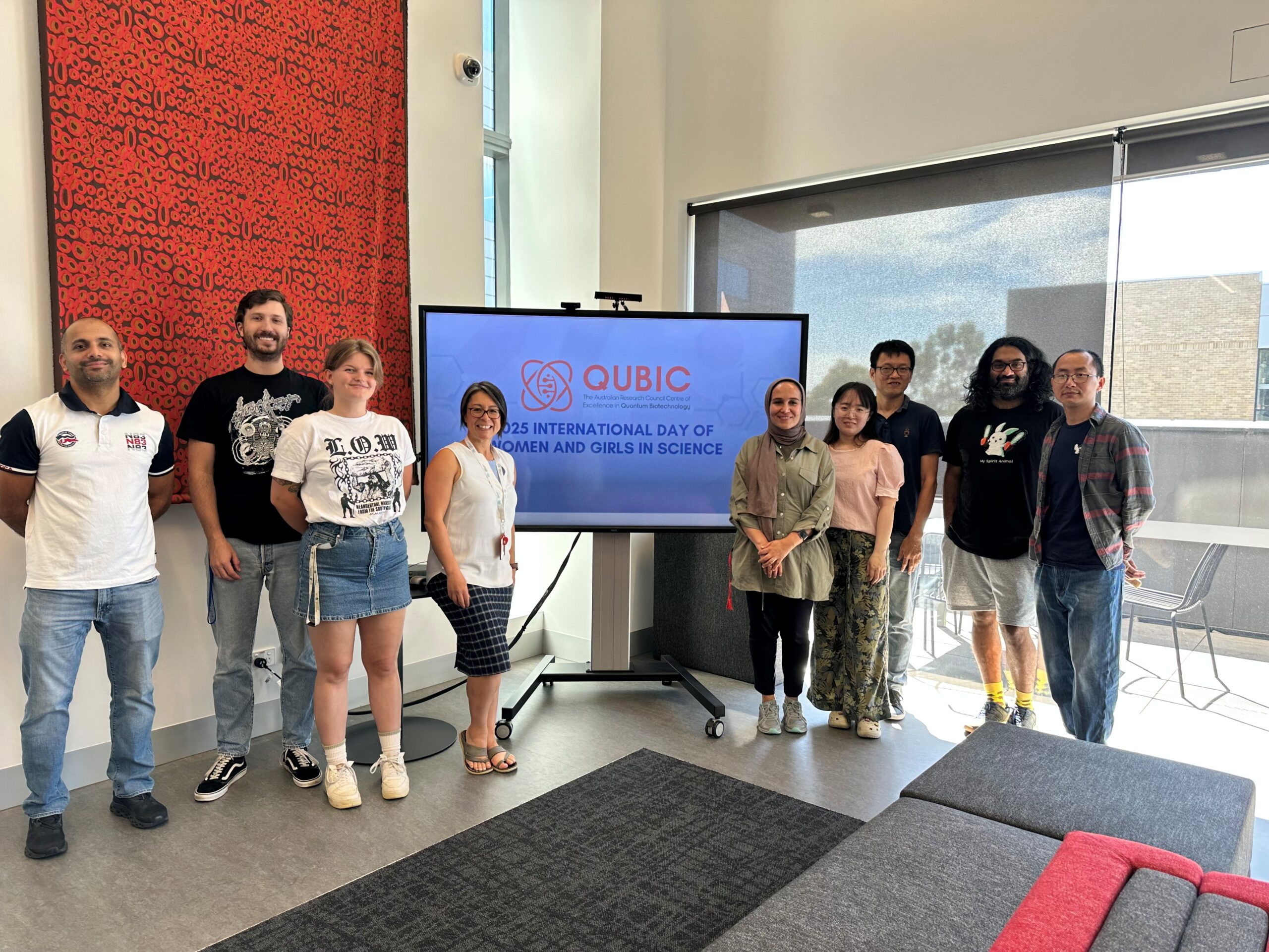
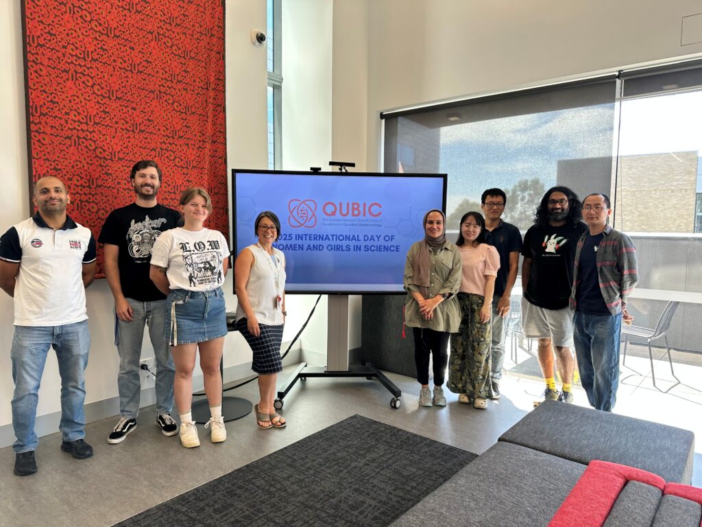
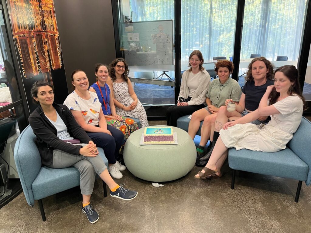
On the UN International Day of Women and Girls in Science, QUBIC proudly celebrated the brilliant women driving innovation in quantum biotechnology. This year’s theme, “Unpacking STEM Careers: Her Voice in Science,” underscores the importance of amplifying women’s contributions and ensuring their voices shape the future of research.
Across our nodes, we organised a variety of events, including morning teas, BBQs, and interactive activities, to celebrate and showcase the achievements of women in science. These events aimed to inspire the next generation of scientists by highlighting the remarkable work of female researchers. It was truly inspiring to hear the many stories of past generations of women scientists and how their journeys and life’s work have significantly contributed to our understanding of the world. Their legacies continue to inspire and pave the way for future generations.
Our latest Researcher Spotlight features one of the many incredible scientists in our community, showcasing her groundbreaking work and dedication to advancing quantum biotechnology. This spotlight is a testament to the talented women who are making significant strides in STEM fields.
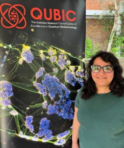
Researcher Spotlight – Dr Nishta Arora
Dr. Nishta Arora, a researcher at QUBIC, is working on cutting-edge technology that could revolutionise brain imaging. Nishta is developing ultrasensitive optomechanical magnetometers for magnetoencephalography (MEG), a technique for mapping brain activity by measuring the magnetic fields produced by neural activity. Her goal? To create compact, room-temperature sensors that make MEG more accessible, portable, and cost-effective.
Her journey in science began with a childhood fascination with physics and engineering, particularly at the micro and nanoscale. Today, she’s translating that curiosity into groundbreaking applications in biomedical sensing.
What she finds most rewarding about being a scientist?
“The ability to push the boundaries of knowledge and create something entirely new is incredibly exciting. I love the process of problem-solving, working with a team of brilliant scientists, and seeing how fundamental research can translate into technologies with real societal impact.”
Her advice to young women considering a career in science?
“Follow your curiosity, and don’t be afraid to take on challenges! Seek mentors, stay persistent, and remember that setbacks are part of the learning process. Most importantly, believe in yourself.”
Let’s continue to champion diverse voices, break down barriers, and inspire the next generation of women in science. Together, we can create a more inclusive and innovative future.
QUBIC’s inaugural Undergraduate Summer Internship Program culminated in a fantastic series of final presentations on Friday 7 February, where the interns showcased the impressive projects they undertook throughout the four-week program.
Developed by QUBIC’s Mentoring, Training, and Development Portfolio (MTD), the program offers the next generation of researchers a valuable hands-on experience in quantum biotechnology research. Participants worked alongside experienced academics in the lab on real-world projects with genuine outcomes.
Alex Wright (left) who is studying a B. Advanced Science / B. Creative Intelligence and Innovation at the University of Technology Sydney focused on the detection and quantification of Amyloid-β biomarkers for Alzheimer’s Disease in plasma, supervised by Chief Investigator A/Prof. Jiajia Zhou.
“I’ve learned so much this past month, especially about the precision required in lab work”, says Alex. “Observing and conducting new processes with an amazing team has been incredibly rewarding. A key takeaway for me is the importance of perfecting protocols to achieve the best results while minimizing costs. A highlight was preparing LFA strips, which was fascinating after using RAT tests during the COVID pandemic without understanding the underlying process.”
Laz Ashcroft (left), a B. Biotechnology student from the University of Wollongong, analysed neuronal function using novel quantum tools with Chief Investigator Professor Lezanne Ooi, and Dr Dzung Do-Ha.
“This internship was a fantastic experience” says Laz. “It has given me a taste of culturing cells, running assays, and analysing results. It boosted my confidence as a scientist, especially with the autonomy to plan and execute experiments. I also enjoyed learning to use GraphPad Prism and improving my presentation skills with feedback from my supervisors. Overall, it was a rewarding and empowering journey.”
The final presentations were inspiring, emphasising the importance of the program in nurturing the up-and-coming generation of scientists in quantum biotechnology. The interns’ dedication and passion for their projects was evident as they shared their findings and reflected on the skills they had learnt throughout the experience. Each presentation was a testament to their hard work, and highlighted the diverse research being conducted:
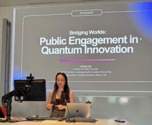
Anthea Sun, UQ – Explored the challenges in public engagement with quantum technology under the guidance of Allison Fish.
Alex Wright, UTS – Focused on the detection and quantification of Amyloid-β biomarkers for Alzheimer’s Disease in plasma, mentored by Jiajia Zhou.
Laz Ashcroft, UOW – Analyzed neuronal function using novel quantum tools, with support from Lezanne Ooi and Dzung Do-Ha.
Nicholas Fantham, UOW – Conducted molecular dynamics simulations of the secondary structure propensities of the conserved region in TDP-43, supervised by Haibo Yu.
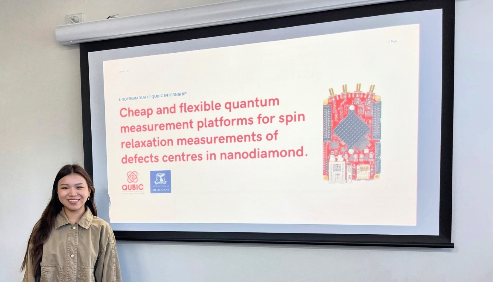
Lianne Lay (above), Uni Melb – Developed cheap and flexible quantum measurement platforms for spin relaxation measurements of defect centers in nanodiamond, guided by David Simpson.
“QUBIC’s inaugural summer internship was an exciting opportunity to engage the next generation of scientists in the rapidly evolving field of quantum biotechnology” said Dr Dzung Do-Ha, program coordinator. “Seeing these bright students explore cutting-edge concepts, develop hands-on skills, and contribute fresh perspectives has been truly inspiring. We are proud to support their growth and look forward to the impact they will make in the future.”
About the Program
The MTD Portfolio Undergraduate Summer Internship Program offers a unique opportunity for current domestic undergraduate students to engage in hands-on quantum biotechnology research projects. Over the course of four weeks, interns gain exposure to the world-class research being conducted within QUBIC laboratories. Hosted by our Chief Investigators at four of QUBIC’s nodes—University of Melbourne, University of Wollongong, University of Queensland, and University of Technology Sydney—the program provides:
Hands-on experience to develop essential research skills
Exposure to cutting-edge research at the interface between quantum technology and biotechnology
Access to world-class researchers and facilities
Networking opportunities with fellow interns and researchers across our nodes
The program will be offered again in 2026. If you would like to know more, please contact program coordinators, Professor Haibo Yu or Dr Dzung Do-Ha.
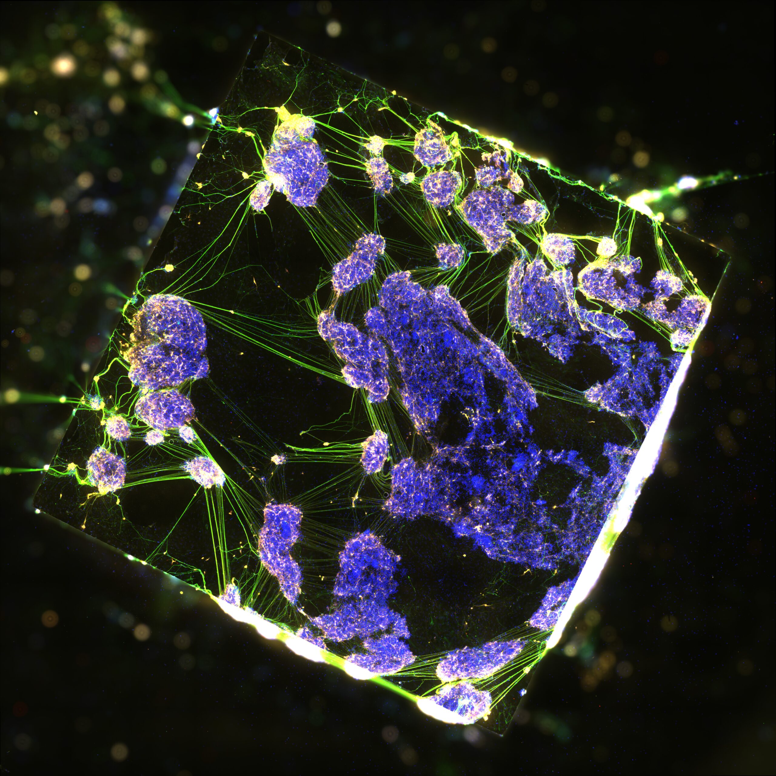
Since the 1970’s the gold standard technique for measuring the activity of individual neurons has been patch clamp, which uses an electrode to record the activity of a single neuron.
However, this measurement destroys the cell, limiting the timeframe of experiments to minutes and can only measure the activity of one neuron at a time.
We are developing novel techniques harnessing the power of quantum technologies to provide new ways to measure brain activity from individual neurons, or whole networks of neurons.
We culture the neurons on lab-grown diamonds to visualise neuronal activity. Through imaging neuronal activity, rather than using electrodes as in patch clamp, the measurements can be taken over long periods of time.
In the future this will allow us to visualise activity across the whole brain and understand how individual neurons connect with each other to coordinate the intricate functions of the brain.
Image: Neurons cultured on a diamond, Ooi Lab, University of Wollongong
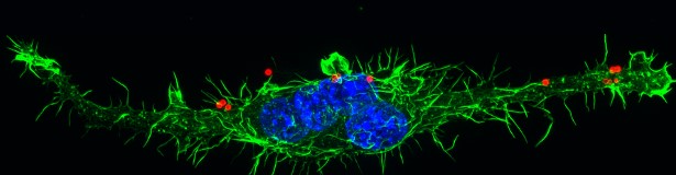
There are many important reasons to look inside bone and the bone marrow buried deep inside bone cavities. Clinicians need to be able to monitor the repair of bone fractures, especially in children and the elderly. Diagnosing and treating bone cancer needs to be guided by tracking the identity and behaviour of cells in bone and marrow. Bone marrow transplants need careful monitoring of the newly-seeded cells. In other tissues within the body, microscopes can be used to help visualize cells and molecules, to understand disease processes, and to guide clinical treatments. In bone, however, this is not currently possible. Bone is a problematic medium for optical microscopes using light or laser sources, due to bone having high light scattering and low penetration properties. The ability to see critical changes in cells or molecules inside bone is severely limited.
Overcoming these current limitations was a challenge set out by the Chan Zuckerberg Initiative (CZI), a philanthropic venture supported by Priscilla Chan and Mark Zuckerberg that seeks to cure human diseases. A team of Australian multi-disciplinary scientists are one of ten teams globally to receive funding through the CZI Deep Tissue Imaging grant to use quantum imaging to overcome the limitations of laser-based imaging and microscopy in bone.
Led by Professor Jennifer Stow, cell biologist at The University of Queensland (UQ) the project will specifically harness innovative quantum triangulation and quantum cutting nanoparticles to image bone marrow using lasers at near infrared wavelengths, in order to create high resolution, deep imaging through bone. Other members of the team are quantum optics scientist Professor Warwick Bowen (UQ); bone marrow expert Professor Allison Pettit (Mater Research); and nanotechnology researchers Distinguished Professor Dayong Jin and Associate Professor Jiajia Zhou at the University of Technology Sydney. The impact of this work is life changing as it will allow researchers and clinicians access to the fine details and diagnostic markers at the cell and molecular level in bone marrow.
Vital expertise and resources for this work comes from the ARC Centre of Excellence in Quantum Biotechnology (QUBIC). The Centre is hosted at The University of Queensland under Director, Professor Warwick Bowen. Other QUBIC Chief Investigators on the team include Stow, Jin and Zhou. Australia’s strength in quantum science, with the support of CZI and its global network, addresses pressing needs for bone marrow imaging in transplantation, cancer and immunity. More broadly, with quantum innovations and training opportunities emerging from this exciting research, we can look forward to the far-reaching benefits for all types of microscopy improving tissue imaging in medicine and biology.
Image: Bone marrow derived immune cells, Stow lab, University of Queensland
 Dr. Nishta Arora, a researcher at QUBIC, is working on cutting-edge technology that could revolutionise brain imaging. Nishta is developing ultrasensitive optomechanical magnetometers for magnetoencephalography (MEG), a technique for mapping brain activity by measuring the magnetic fields produced by neural activity. Her goal? To create compact, room-temperature sensors that make MEG more accessible, portable, and cost-effective.
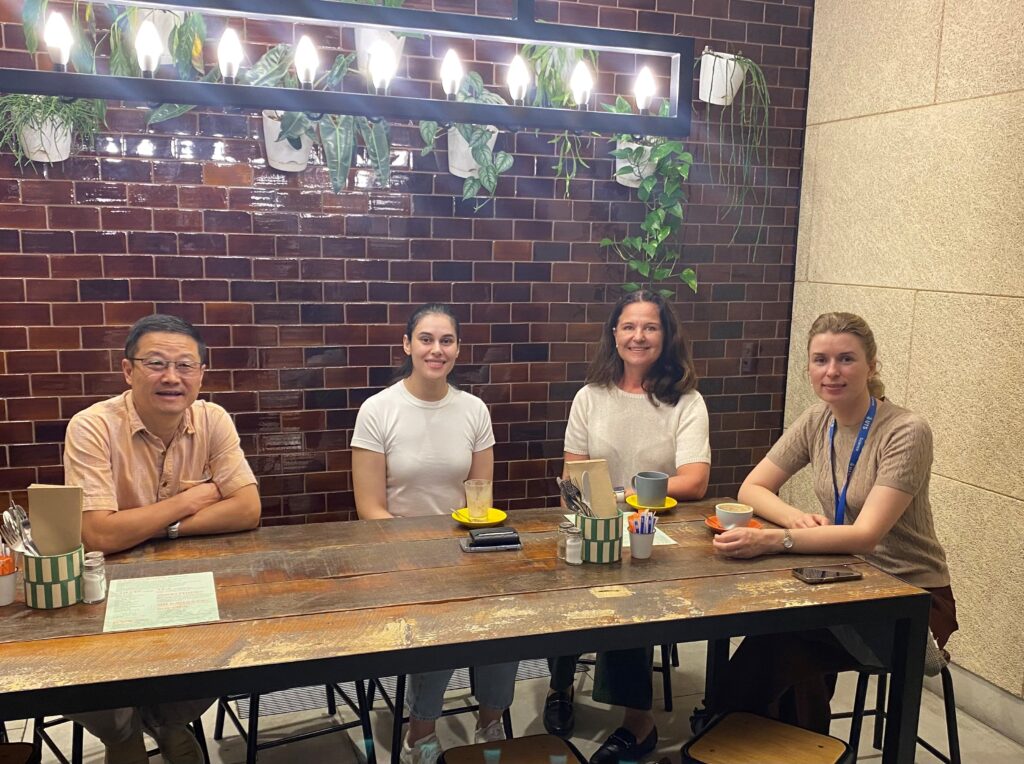
 Alex Wright (left) who is studying a B. Advanced Science / B. Creative Intelligence and Innovation at the University of Technology Sydney focused on the detection and quantification of Amyloid-β biomarkers for Alzheimer’s Disease in plasma, supervised by Chief Investigator A/Prof. Jiajia Zhou.
 Laz Ashcroft (left), a B. Biotechnology student from the University of Wollongong, analysed neuronal function using novel quantum tools with Chief Investigator Professor Lezanne Ooi, and Dr Dzung Do-Ha.